|
|
||
| HOME | ||
|
|
Training Program |
|
|
|
Contact Info |
|
|
|
Training Videos | |
|
|
Atlas of Ultrasound | |
|
|
Hands-on Classes | |
|
|
Bookings | |
|
|
||
|
|
||
|
|
||

|
ARCHIVE |
|||
|
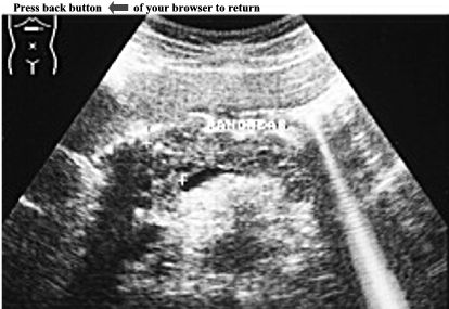
CASE 7: Ultrasound examination of gallbladder showed an asymmetric thickening of the wall (7mm) with a single central hypoechoic zone sandwiched with two echogenic layers, see white arrows. An hyperechoic, round mass (7mm) with posterior acoustic shadow was demonstrated impacted in the neck of the gallbladder (see black arrow). No pericholecystic fluid collection was present. The rest of the examination was normal. ANSWER: Acute Calculous Cholecystitis If you want more information about Acute Calculous Cholecystitis click here. |
|
||

